
二手奥林巴斯(Olympus)BX43 生物显微镜
产品名称: 二手奥林巴斯(Olympus)BX43 生物显微镜
英文名称: OLYMPUS BX43 Biological Microscope
产品编号: BX43
产品价格: 56000
产品产地: 美国
品牌商标: 奥林巴斯
更新时间: 2026-05-14T16:53:20
使用范围: null
- 联系人 : 陈经理
- 地址 : 上海市浦东新区金桥开发区置业路111号3号楼
- 邮编 : 201299
- 所在区域 : 上海
- 电话 : 137****0200 点击查看
- 传真 : 点击查看
- 邮箱 : sales@yinzhisci.com
- 二维码 : 点击查看
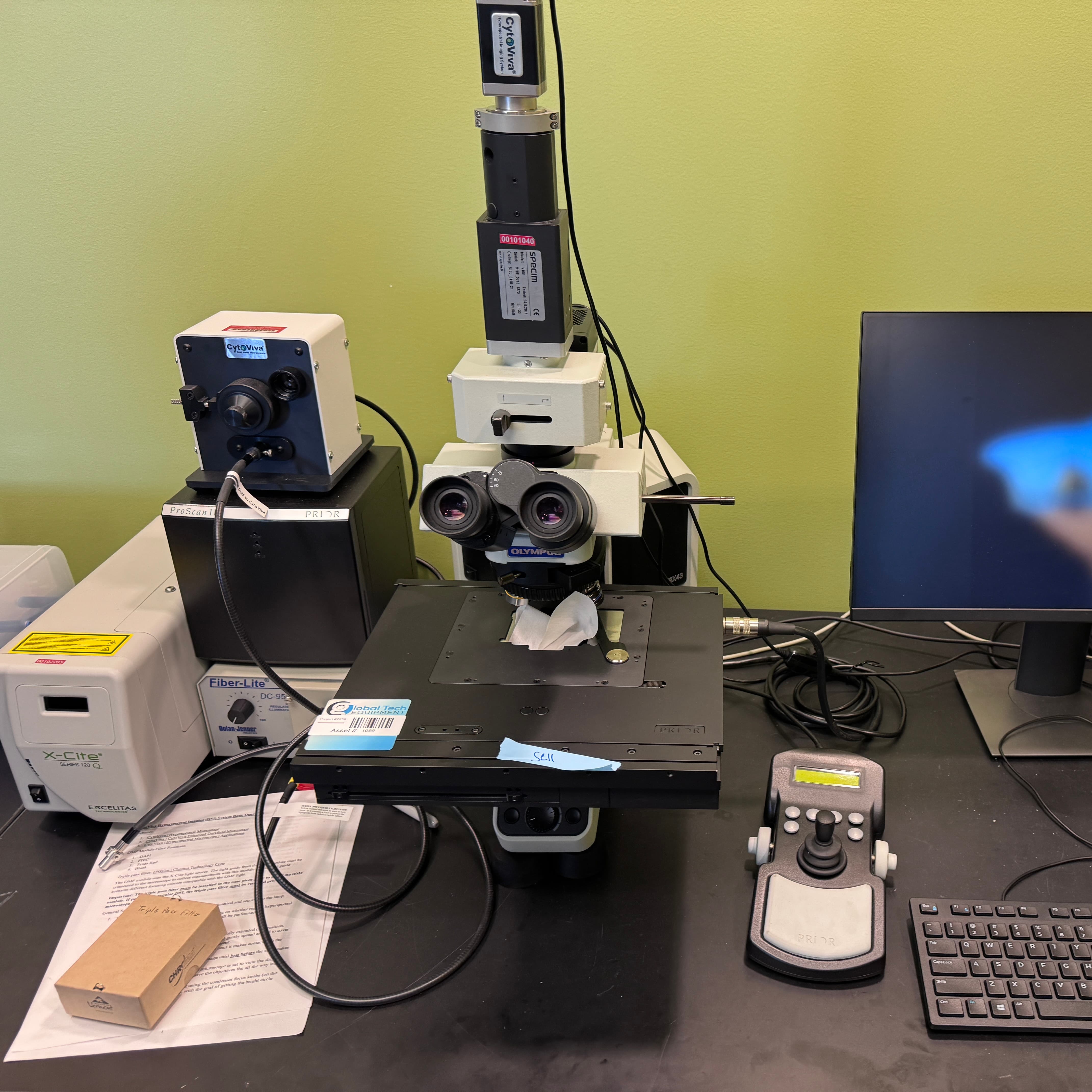
The OLYMPUS BX43 Biological Microscope system, as described with its specialized attachments—including the Specim V10E, CytoViva Dual Mode fluorescence, X-Cite Series 120 Q, Fiber Lite Illuminator, PRIOR ProScan III, PRIOR JoyStick, and a Dell PC with software and dongle—represents a highly sophisticated, research-grade multimodal imaging workstation. Far beyond a standard light microscope, this integrated platform is engineered to tackle complex challenges in cell biology, pathology, nanomedicine, and materials science, offering a unique combination of high-sensitivity fluorescence, label-free nanoscale imaging, and chemical component analysis. At its core, the Olympus BX43 is a modular, ergonomic, and optically superior positive microscope. It utilizes Olympus’s renowned UIS2 (Universal Infinity-corrected Optical System) which delivers exceptional image flatness, high resolution, and minimal chromatic aberration across the entire field of view. The BX43 supports a wide array of contrast techniques, including brightfield, phase contrast, Differential Interference Contrast (DIC), polarization, and darkfield, making it inherently versatile for observing unstained living cells, tissue sections, and material samples. Its robust frame and precision optics provide the stable foundation necessary for the advanced illumination and detection modules mounted upon it.
The illumination and excitation suite of this system is both powerful and precise. The X-Cite Series 120 Q is a 120-watt metal halide arc lamp house, serving as the primary fluorescence excitation source. Renowned for its long bulb life (up to 2,000 hours), stable intensity output, and broad-spectrum emission, it provides consistent and intense light for exciting a wide range of fluorophores (such as DAPI, FITC, TRITC, and far-red dyes) without the rapid decay and warm-up issues associated with traditional mercury vapor lamps. The "Q" (Quiet) model features an internal shutter and adjustable aperture, allowing for precise control of light exposure to minimize photobleaching and phototoxicity in sensitive live-cell or prolonged time-lapse experiments. Complementing this, the Fiber Lite Illuminator (typically a Dolan-Jenner or similar brand industrial fiber optic light source) provides high-intensity, cool, halogen or LED-based transmitted or reflected light for brightfield, darkfield, or general illumination tasks, ensuring even lighting and preventing heat damage to the sample. These light sources feed into the BX43’s optimized fluorescence light path, which often employs a fly-eye lens design to ensure exceptionally uniform illumination across the field of view—a critical requirement for quantitative fluorescence analysis and multi-area scanning.
Where this system truly distinguishes itself is through the integration of the CytoViva Dual Mode fluorescence module and the Specim V10E hyperspectral imaging attachment. The CytoViva Dual Mode system is a proprietary, enhanced illumination technology that replaces the standard condenser. It enables the simultaneous or rapid-toggle observation of traditional wide-field fluorescence and enhanced darkfield (scattering-based) imaging. The groundbreaking advantage of this module is its ability to visualize unstained or "label-free" nanoparticles, viral particles, exosomes, and sub-cellular structures down to approximately 20–100 nm in size, which are typically invisible under standard brightfield or even darkfield microscopy. This allows researchers to track the uptake, distribution, and interaction of nanocarriers (like liposomes or gold nanoparticles) within live cells or tissue slices without relying solely on fluorescent tags, which can sometimes alter particle behavior or quench. Furthermore, it retains full compatibility with standard fluorescent protein (e.g., GFP, RFP) or dye labeling, permitting the co-localization of labeled organelles or proteins with unlabeled nanomaterials in real-time. This dual-modality is revolutionary for fields like nanotoxicology, drug delivery, and viral pathogenesis.
Elevating the system further, the Specim V10E is a push-broom hyperspectral imaging (HSI) camera module that integrates seamlessly with the microscope’s trinocular port or camera mount. Unlike a standard RGB camera that captures only three broad color channels, the V10E acquires a full, continuous spectrum (typically in the range of 400–1000 nm, covering Visible to Near-Infrared) for every single pixel in the field of view. This "hypercube" of data (x, y, and λ) enables "spectroscopic imaging," where the user can not only see the morphology of the sample but also identify and map the spatial distribution of different chemical components based on their unique spectral "fingerprints." For example, it can differentiate between various fluorescent dyes, distinguish autofluorescence from specific staining, quantify drug distribution within tissues, or identify mineral compositions in geological samples, all without destructive testing. This turns the microscope into a powerful analytical instrument for quantitative, non-invasive chemical mapping.
To manage these advanced imaging modalities and translate them into actionable data, the system relies on a fully automated control and digital workflow ecosystem. The PRIOR ProScan III is a high-precision, closed-loop, motorized XY stage controller, paired here with a PRIOR JoyStick for intuitive manual navigation. The ProScan III offers sub-micron repeatability and programmable movement, enabling automated functions such as large area tiling (stitching multiple adjacent fields together to create a single high-resolution overview image of a large tissue section), multi-point time-lapse imaging, Z-stack acquisition for 3D reconstruction, and high-throughput screening of multiple samples on a slide or multi-well plate. The inclusion of a Dell PC, pre-loaded with dedicated microscope control and image analysis software (such as Olympus cellSens, Prior’s StagePro, CytoViva’s ENVI software, and Specim’s Lumo Recorder/Analyzer), along with the necessary hardware dongle, ensures that the operator has a turnkey, licensed environment to control all hardware parameters (light sources, shutter, stage movement, Z-focus, filter wheels), acquire images, perform hyperspectral unmixing, conduct quantitative fluorescence measurements, and generate comprehensive reports.
In summary, this specific configuration of the Olympus BX43 is not merely a tool for looking at cells; it is a comprehensive, multimodal nano-bioimaging and analytical workstation. It empowers researchers to conduct routine fluorescence assays, perform cutting-edge single-particle tracking of nanomaterials in biological matrices, and execute spatially resolved chemical characterization of complex heterogeneous samples, all within a single, integrated, and automated platform. This makes it an invaluable asset for advanced life science research labs, core imaging facilities, and multidisciplinary material science centers.
Key Technical Components & Specifications:
|
Component |
Key Features / Specifications |
|---|---|
|
Core Microscope |
Olympus BX43 Positive Biological Microscope; UIS2 Infinity Optical System; Supports BF, PH, DIC, FL, DF; Trinocular Tube. |
|
Hyperspectral Imager |
Specim V10E; Spectral Range 400–1000 nm (Visible-NIR); Push-broom line scan; High spectral resolution (approx. 2–5 nm). |
|
Dual Mode Module |
CytoViva Dual Mode Fluorescence/Darkfield; Enhanced illumination; Detects label-free nanoparticles down to ~20–100 nm. |
|
FL Excitation Source |
X-Cite Series 120 Q; 120W Metal Halide; >2000 hr lamp life; Stable intensity; Internal shutter. |
|
Transmitted Illuminator |
Fiber Lite Illuminator; Fiber optic light guide; High-intensity halogen/LED; Cool operation. |
|
Motorized Stage |
PRIOR ProScan III; Closed-loop XY; Sub-micron accuracy; Programmable scanning (Tiling, Z-stack, Time-lapse). |
|
Stage Controller |
PRIOR JoyStick; Ergonomic manual control of ProScan III stage. |
|
Control PC & Software |
Dell Workstation; Microscope control software (e.g., cellSens, StagePro); Hyperspectral software (Lumo); CytoViva software; Hardware Dongle for licensing. |
|
Typical Applications |
Nanoparticle-cell interactions, Drug delivery, Viral entry, Immunofluorescence, Tissue pathology, Chemical mapping, Live-cell imaging. |